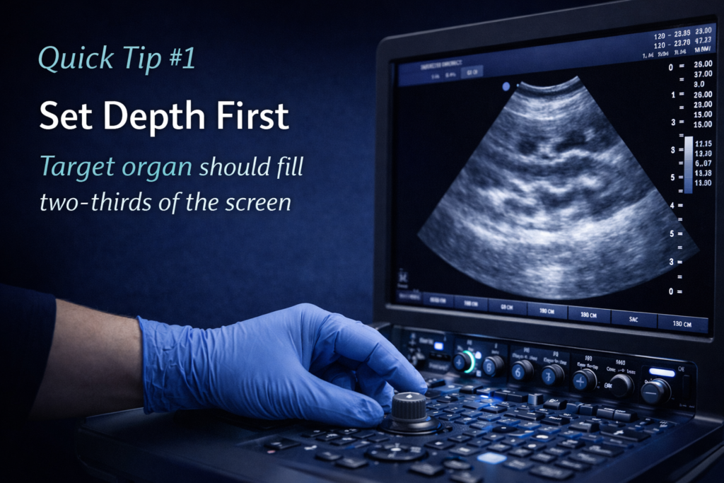
Improve Image Quality by Adjusting Depth First
One of the fastest ways to improve your veterinary ultrasound image is to optimise depth before changing anything else. A common mistake is scanning too deep, leaving the area of interest too small on the screen and reducing detail.
Try this simple rule
Set your depth so the target organ fills approximately two-thirds of the screen.
This allows:
- better resolution
- improved anatomical detail
- easier lesion detection
- more confident measurements
- cleaner cine loops and saved images
For example:
- liver and gallbladder → reduce unnecessary far field depth
- bladder → keep the apex and neck centred
- kidneys → fill the screen without excess surrounding tissue
- echo → optimise depth so chambers occupy the image appropriately
Why it matters
Depth is often the first knobology adjustment that immediately sharpens confidence. Before touching gain, ask: Am I simply scanning too deep?
Small adjustments create better images.
Altitude810 Tip: Optimise depth first, then fine-tune gain and focal zones.